Urticaria: An Evidence-Based Clinical Guide
Introduction
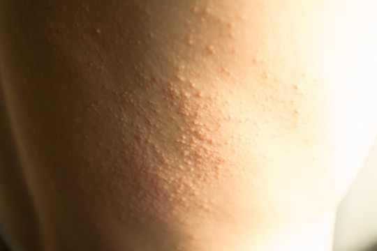
Urticaria, commonly known as hives, is characterized by transient, pruritic wheals and/or angioedema resulting from localized plasma extravasation in the dermis. Lesions typically blanch with pressure, vary in size, and resolve within 24 hours without residual pigmentation. While acute urticaria often follows identifiable exposures, chronic urticaria—defined by daily or almost daily symptoms persisting beyond six weeks—can profoundly impact quality of life and warrants systematic evaluation and management.
Epidemiology and Classification
- Incidence & Prevalence: Affects up to 20% of the general population at some point; chronic spontaneous urticaria (CSU) occurs in ~1% of adults, with a female predominance (2:1).
- Age Distribution: Acute urticaria spans all ages; CSU peaks between 30–50 years. Pediatric urticaria accounts for 15–25% of childhood rash presentations.
- Classification:
- Acute Urticaria: Symptoms <6 weeks; often IgE-mediated (foods, drugs, insect stings), viral infections, or idiopathic.
- Chronic Urticaria: ≥6 weeks. Subtypes include:
- Chronic Spontaneous Urticaria (CSU): No external trigger identified.
- Chronic Inducible Urticaria (CIndU): Physical triggers (dermographism, cold, heat, pressure, cholinergic).
- Angioedema: Deep dermal/subcutaneous edema, may occur with or without urticarial wheals.
Pathophysiology
- Mast Cell Activation: Central event in urticaria; degranulation releases histamine, leukotrienes, prostaglandins, and cytokines.
- IgE-Mediated Mechanisms: Allergen cross-linking on FcεRI receptors triggers immediate response.
- Non–IgE Pathways: Autoimmune (anti–IgE or anti-FcεRI antibodies), complement activation, direct mast cell degranulators (opioids, radiocontrast).
- Physical Factors: Mechanical, temperature, or cholinergic stimuli induce localized mast cell mediator release.
Clinical Evaluation
History
- Onset & Duration: Timing relative to exposures, seasonal or continuous pattern.
- Morphology & Distribution: Wheal size, central pallor, dermographism.
- Associated Symptoms: Pruritus severity, angioedema (facial, laryngeal), systemic signs (fever, arthralgia).
- Precipitating Factors: Recent infections, new medications, foods, environmental exposures, stress.
- Past Medical History: Autoimmune disease, thyroid dysfunction, atopic disorders.
Physical Examination
- Skin Exam: Document transient nature of wheals; positive dermographism test if stroking skin with blunt object produces a linear wheal.
- Angioedema Assessment: Check periorbital, lip, tongue, and laryngeal regions.
- Systemic Evaluation: Vitals, joint exam, lymphadenopathy (to exclude vasculitis or systemic disease).
Differential Diagnosis
- Urticarial vasculitis (lesions last >24 hours, burn or sting rather than itch, residual hyperpigmentation, biopsy shows leukocytoclastic vasculitis).
- Mastocytosis (Darier’s sign, systemic symptoms).
- Autoinflammatory syndromes (cryopyrin-associated periodic syndromes).
- Fixed drug eruption (persistent, well-demarcated plaque at same site).
Diagnostic Workup
- Targeted Laboratory Tests for CSU: CBC with differential, ESR/CRP, thyroid-stimulating hormone (TSH) and antithyroid antibodies, hepatitis serologies when risk factors present.
- Infectious Triggers: Consider viral panels (e.g., parvovirus B19), Helicobacter pylori testing in refractory cases.
- Autoimmune Screening: ANA, complement levels if vasculitis suspected.
- Skin Biopsy: Reserved for atypical or vasculitic presentations.
Management
First-Line Therapy
- Second-Generation H1-Antihistamines: Nonsedating agents (cetirizine, loratadine, fexofenadine) at standard doses; well tolerated, once daily.
- Ultrapotent H1-Updosing: If symptoms persist after 2–4 weeks, increase dose up to 4× standard dose.
Adjunctive and Second-Line Options
- H2-Antagonists: Ranitidine or famotidine added to H1 blockade for refractory pruritus.
- Leukotriene Receptor Antagonists: Montelukast in selected patients with NSAID-sensitive CSU.
- Omalizumab: Anti-IgE monoclonal antibody; indicated for CSU unresponsive to high-dose antihistamines.
- Cyclosporine: 3–5 mg/kg/day for severe, refractory cases; monitor renal function and blood pressure.
Acute Severe Reactions
- Anaphylaxis Protocol: Epinephrine intramuscularly, airway management, IV fluids, corticosteroids, and H1/H2 antihistamines.
- Angioedema without Urticaria: Evaluate for C1-INH deficiency; treat with C1 inhibitor concentrate or icatibant in hereditary angioedema.
Patient Education and Support
- Trigger Avoidance: Maintain an allergy diary to identify and eliminate causative factors.
- Lifestyle Modifications: Stress reduction, warm showers (avoid temperature extremes), loose clothing.
- Medication Adherence: Counsel on dose escalation and duration; discuss side effect profiles.
- Quality of Life: Assess impact using validated tools (e.g., Urticaria Activity Score, Chronic Urticaria Quality of Life Questionnaire).
Special Populations
- Pediatric Considerations: Dosage adjustments, safety of omalizumab in children ≥6 years, monitor growth parameters.
- Pregnancy and Lactation: Loratadine and cetirizine considered safe; avoid first-generation sedating antihistamines unless necessary.
Prognosis and Follow-Up
- Acute Urticaria: Most resolve within days to weeks once trigger removed.
- Chronic Urticaria: Median duration 1–5 years; spontaneous remission in ~50% by year 1, 80% by year 5.
- Monitoring: Reassess every 3–6 months; taper therapy once symptom-free for 3 months.
Conclusion
Effective management of urticaria hinges on precise classification, identification of reversible triggers, and a stepwise therapeutic approach. Emerging biologics and immunomodulators offer hope for refractory cases, while patient education and support remain fundamental to improving outcomes and quality of life.


