Skin Cancer Etiology and Risk Stratification
Introduction
Skin cancer comprises melanoma and non‑melanoma skin cancers (basal cell carcinoma and cutaneous squamous cell carcinoma). Ultraviolet (UV) radiation is the dominant causal exposure, with additional roles for ionizing radiation, chemical carcinogens, chronic inflammation/scar tissue, and host susceptibility. Early risk recognition enables targeted prevention and timely biopsy of suspicious lesions.
Major Causes and Risk Factors
1) Ultraviolet radiation (UVB and UVA)
– Cumulative and intermittent high‑intensity sun exposure drives DNA damage (cyclobutane pyrimidine dimers, 6‑4 photoproducts) and oxidative stress.
– 80% of NMSC arises on sun‑exposed sites (face, scalp, ears, neck, forearms, dorsal hands).
2) Ionizing and thermal radiation
– Occupational/medical exposure (e.g., radiation workers, prior radiotherapy fields) increases NMSC risk within irradiated skin.
3) Genetic and pigmentary disorders
– Xeroderma pigmentosum (defective nucleotide excision repair) and oculocutaneous albinism markedly elevate risk.
– Fair skin (Fitzpatrick I–II), red/blond hair, freckling, MC1R variants confer higher UV susceptibility.
4) Chemical exposures
– Chronic contact with arsenic; polycyclic aromatic hydrocarbons in tar, pitch, asphalt, petroleum derivatives.
5) Chronic skin injury and inflammation
– Long‑standing scars, burns, sinus tracts, and non‑healing ulcers can transform—classically to cSCC (Marjolin ulcer).
6) Host and demographic factors
– Male sex, age >50, immunosuppression (solid‑organ transplant, hematologic malignancy), prior skin cancer, numerous/atypical nevi.
Pathogenesis: How Damage Becomes Cancer
- Direct genotoxicity: UV‑induced DNA photoproducts create signature C→T transitions when unrepaired.
- Oxidative stress and inflammation: ROS generation, immunosuppression, and clonal expansion of mutated keratinocytes/melanocytes.
- Field cancerization: widespread actinic damage predisposing to multifocal NMSC in sun‑exposed areas.
Clinical Risk Stratification (at a glance)
- High risk: immunosuppressed; history of multiple NMSC; genetic repair disorders; heavy outdoor/indoor tanning exposure; prior radiation field; rapidly growing or recurrent lesions; high‑risk anatomic sites (mask areas of face, ears, lips, scalp).
- Intermediate risk: fair skin with significant cumulative sun exposure; numerous actinic keratoses; age >65.
- Lower risk: darker skin types without additional risk factors (note: melanoma can still occur, often on acral or mucosal sites).
Prevention and Counseling
- Photoprotection: shade seeking; protective clothing (UPF), wide‑brim hats, UV‑blocking sunglasses; broad‑spectrum SPF ≥30 sunscreen applied generously and reapplied every 2 hours and after swimming/sweating.
- Behavioral: avoid midday sun; eliminate indoor tanning; plan sun‑safe travel and outdoor work routines.
- Environment/occupational: minimize chronic contact with tar/asphalt/petroleum derivatives; adhere to radiation safety protocols.
- Chemoprevention: consider nicotinamide 500 mg twice daily in high‑risk patients for NMSC reduction (adjunct to photoprotection).
- Education: monthly skin self‑exams; teach ABCDE for pigmented lesions and the “ugly duckling” sign; photograph lesions for comparison.
When to Biopsy or Refer Urgently
- New or evolving pigmented lesion (asymmetry, border irregularity, color variegation, diameter ≥6 mm, or evolving).
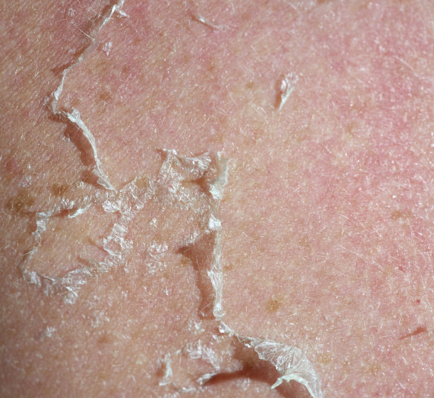
- Firm, growing nodule; non‑healing ulcer on sun‑exposed skin; tender indurated plaque.
- Lesion with perineural symptoms (pain, numbness) or arising in a chronic scar/ulcer.
- Suspicion for melanoma or high‑risk cSCC/BCC in cosmetically/functional critical sites (consider Mohs or excisional biopsy).
Key Takeaways
- UV exposure is the principal modifiable driver; genetics, chemicals, radiation, and chronic wounds add risk.
- Systematic risk assessment plus photoprotection and patient education prevent many NMSC cases and aid earlier melanoma detection.
- Maintain a low threshold for biopsy of suspicious or changing lesions, especially in high‑risk patients and sites.