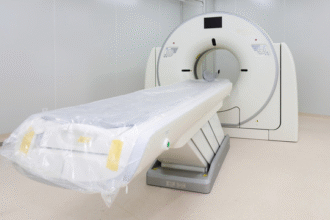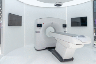
MRI Advantages, Safety Considerations, and Patient Preparation
Magnetic Resonance Imaging (MRI) has matured into a core diagnostic platform since the first clinical head images were produced in the late 1970s. Its evolution has paralleled advances in superconducting magnet engineering, gradient performance, multichannel radiofrequency (RF) coil design, parallel acquisition strategies, and sophisticated reconstruction algorithms. Unlike ionizing modalities, MRI derives contrast from intrinsic nuclear magnetic properties—principally hydrogen proton density and relaxation dynamics—supplemented by motion, diffusion, susceptibility, perfusion, and chemical shift phenomena. This multi-parametric versatility allows a single examination to capture complementary structural, functional, and in selected contexts metabolic or molecular information, substantially reducing diagnostic uncertainty across neurologic, musculoskeletal, cardiovascular, abdominal, pelvic, and oncologic domains.
The principal advantages of MRI stem from its superior soft tissue contrast, multiplanar native acquisition without geometric distortion, and absence of ionizing radiation. Brain, spinal cord, marrow, ligament, cartilage, pelvic organ, prostate zonal, and myocardial tissue characterization exceed that of computed tomography in most non-calcific contexts. MRI’s ability to tailor contrast via pulse sequence manipulation enables targeted evaluation of edema, acute ischemia (diffusion restriction), demyelination, hemorrhagic byproducts, fibrosis, or neoplastic angiogenesis. Advanced techniques—diffusion tensor imaging, functional MRI (BOLD), dynamic contrast perfusion, MR spectroscopy, T1/T2 mapping, quantitative susceptibility mapping, arterial spin labeling, and whole-body diffusion—extend capability into microstructural integrity assessment, cortical activation mapping, hemodynamic profiling, metabolic fingerprinting, and systemic disease staging without incremental radiation burden. These attributes are particularly valuable in longitudinal surveillance of pediatric patients, reproductive-age individuals, and oncology survivors where cumulative radiation is a concern.
MRI safety is governed by four interacting physical domains: the static magnetic field (B₀), time‑varying gradient fields, RF energy deposition (specific absorption rate, SAR), and acoustic noise. The static field exerts translational and rotational forces on ferromagnetic objects, creating projectile risks and potential device displacement or torque. Time‑varying gradients can induce peripheral nerve stimulation and, in rare circumstances, audible vibration discomfort. RF absorption increases tissue temperature, managed through adherence to SAR regulatory limits and appropriate patient screening (fever, impaired thermoregulation). Gradient coil vibration produces high sound pressure levels; acoustic protection with earplugs or headphones is mandatory. Comprehensive pre-scan screening mitigates the majority of preventable adverse events.
Implant and foreign body assessment requires nuanced classification rather than oversimplified absolute exclusion. Many modern vascular stents, orthopedic internal fixation constructs, and numerous aneurysm clips are labeled MR conditional, permitting scanning within specified field strengths, spatial gradient limits, and SAR constraints. Legacy ferromagnetic aneurysm clips without documentation, certain older pacemakers and implantable cardioverter-defibrillators (ICDs), retained intraorbital metallic fragments (especially in metal workers), and some neurostimulators necessitate either alternative imaging, ex vivo artifact testing, device interrogation, or supervised conditional protocols with cardiology or electrophysiology support. Contemporary MR conditional cardiac rhythm devices can be safely imaged under standardized programming and monitoring. Intrauterine devices composed of non-ferromagnetic alloys or polymers generally pose no hazard, though artifact extent should be anticipated in pelvic imaging. Titanium, being paramagnetic and weakly interacting with B₀, neither migrates nor produces substantial susceptibility distortion except in high-field gradient echo sequences. Sharp fragment proximity to critical neurovascular structures (e.g., intraocular) remains a relative contraindication until excluded by preliminary radiography or CT.
Patient preparation emphasizes removal of external metallic objects and electronics to prevent projectile events, localized heating, demagnetization, or image artifact. This includes jewelry, piercings, watches, wallets with magnetic stripes, hearing aids, insulin pumps, continuous glucose monitors, ECG leads with ferromagnetic clips, and cosmetic items containing metallic particles (certain eye makeup). Clothing should be free of metallic fibers or reflective threads that may concentrate RF energy. Thorough verbal briefing reduces anxiety-induced motion; anxiolysis or monitored sedation may be considered for severe claustrophobia. Pediatric, cognitively impaired, or critically ill patients benefit from tailored immobilization, physiologic monitoring, and abbreviated or motion-robust protocol design using single-shot, parallel, and compressed sensing acquisitions. Clear communication regarding breath-holds, stillness, and noise expectation improves success rates and reduces need for rescanning.
While MRI excels in soft tissue evaluation, its limitations inform modality selection. Scan durations can challenge unstable or noncompliant patients; emergent assessment of major trauma, acute intracranial hemorrhage triage, or suspected pneumothorax remains the domain of CT for speed and universal availability. Cortical bone and fine calcification are incompletely characterized due to low proton density and rapid signal decay, though ultrashort echo time (UTE) and zero echo time (ZTE) techniques are narrowing this gap. Susceptibility to motion (respiratory, cardiac, peristaltic) can degrade abdominal and thoracic imaging, mandating gating, motion correction, or rapid sequence substitutions. Device-related artifacts (magnetic susceptibility, RF shielding, geometric distortion) may obscure adjacent anatomy. Cost, scheduling constraints, and geographic access inequities further temper widespread immediate deployment despite high diagnostic value.
Gadolinium-based contrast agents (GBCAs) expand lesion conspicuity, vascular delineation, and blood–brain barrier integrity assessment by shortening T1 relaxation times in regions of extracellular distribution. Their administration is generally safe; allergic-like reactions are uncommon and usually mild. Risk mitigation includes renal function screening to avoid administration in patients with advanced chronic kidney disease (particularly for linear non‑ionic agents historically associated with nephrogenic systemic fibrosis). Macrocyclic agents with higher kinetic stability are preferred when renal reserve is marginal. Emerging concerns regarding gadolinium deposition in the brain and bone have not demonstrated definitive clinical harm but support a stewardship approach—administering the lowest effective dose and reserving repeat contrast studies for altering management.
Emergent innovation continues to refine MRI’s risk–benefit balance. Parallel transmit technology improves B₁ field homogeneity at higher field strengths, reducing dielectric shading and enabling more uniform abdominal and cardiac imaging at 3 Tesla and beyond. Deep learning reconstruction integrates learned priors to preserve edge detail while suppressing noise, restoring diagnostic confidence at accelerated acquisition factors previously unattainable with conventional parallel imaging alone. Quantitative mapping sequences (T1, T2, T2*, proton density fat fraction) are progressing from research to routine clinical biomarker status, enabling standardized longitudinal assessment of diffuse myocardial fibrosis, liver iron overload, hepatic steatosis, and neurodegenerative microstructural change. MRI-guided focused ultrasound offers non-invasive therapeutic interventions including tremor ablation, targeted blood–brain barrier modulation for drug delivery trials, and investigational oncologic applications. Hybrid PET/MR platforms synergize metabolic and high-resolution soft tissue imaging, particularly in neuro-oncology, head and neck malignancy, and pediatric lymphoma staging, reducing cumulative radiation dose compared with PET/CT.
In totality, MRI leverages a confluence of physics, engineering, and computational strategy to deliver rich, multi-dimensional insights into human anatomy and pathology without ionizing exposure. Its safe and effective use hinges on rigorous screening, informed device evaluation, protocol customization, artifact mitigation, and judicious contrast deployment. As acquisition acceleration, quantitative reproducibility, and intelligent automation mature, MRI’s role will further expand in precision diagnostics, phenotypic disease stratification, and image-guided therapy while reinforcing patient-centered safety principles.
Disclaimer: Educational synthesis; defer to institutional MRI safety officer and published professional society guidelines (e.g., ACR, ISMRM) for definitive protocol governance.