Clinical Guide for Cortical Cataract
Introduction
Cortical cataract is the most common subtype of age-related lens opacification, characterized by opacities in the lens cortex that progress from peripheral vacuoles to dense wedge-shaped spokes. While early stages may be asymptomatic, advancing cortical cataracts impair visual acuity, contrast sensitivity, and quality of life. This guide reviews epidemiology, pathophysiology, clinical staging, diagnostic evaluation, management strategies, and postoperative care to support evidence-based practice.
Epidemiology and Risk Factors
- Prevalence: Age-related cortical cataract prevalence increases from <5% in individuals aged 50–59 years to >50% in those over 80.
- Risk Factors:
- Age: Principal determinant; cumulative lens protein glycation and oxidative stress.
- Ultraviolet (UV) Exposure: UV-B radiation induces cortical fiber damage.
- Metabolic Disorders: Diabetes mellitus accelerates osmotic stress within lens fibers.
- Medications: Chronic corticosteroids contribute to cataractogenesis.
- Genetic Predisposition: Polymorphisms in crystallin genes may influence susceptibility.
Pathophysiology and Classification
Pathogenesis
Cortical cataract formation begins with disruption of lens fiber cell membrane integrity, leading to localized hydration (vacuole formation) and protein aggregation. Progressive cortical dehydration and calcification advance opacification, altering refractive index and increasing light scatter.
Clinical Staging
- Incipient (Initial) Stage:
- Peripheral cortical vacuoles and hydrops develop; lamellar separation appears.
- Visual acuity usually remains intact unless opacities encroach pupillary axis.
- Immature (Expansion) Stage:
- Wedge-shaped spokes radiate toward the center; cortical opacities merge.
- Lens hydration increases, anterior chamber shallows; risk of phacomorphic glaucoma.
- Mature Stage:
- Complete cortical opacification; nucleus clear or mixed; pupillary reflex absent.
- Vision typically reduced to hand motion or light perception; fundus view obscured.
- Hypermature (Overmature) Stage:
- Cortical liquefaction (cortical emulsification) and lens shrinkage; nucleus may sink (Morgagnian cataract).
- Iris tremor and darkened anterior chamber noted; visual acuity variable.
Clinical Evaluation
History
- Symptom Onset & Progression: Blurred vision, glare (especially in bright light or night driving), monocular diplopia.
- Associated Symptoms: Photophobia, difficulty reading, color desaturation.
- Risk Assessment: UV exposure, systemic diseases (e.g., diabetes), medication history, prior ocular inflammation or trauma.
Examination
- Visual Acuity & Refraction: Assess best-corrected visual acuity (BCVA) and measure refractive change; cortical cataracts often induce hyperopic shift initially.
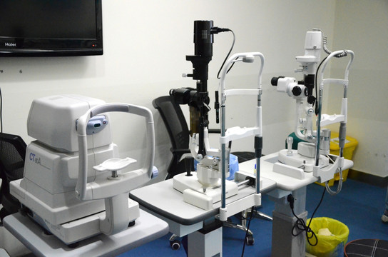
- Slit-Lamp Biomicroscopy: Document cortical spokes, vacuoles, zonular integrity, anterior chamber depth.
- Tonometry: Measure intraocular pressure; shallow chamber in immature stages suggests phacomorphic risk.
- Fundus Examination: Dilated exam when media clarity permits; retinal evaluation influences surgical planning.
Differential Diagnosis
- Nuclear sclerotic cataract (central yellow–brown nucleus)
- Posterior subcapsular cataract (PSC; affects near acuity)
- Secondary cataracts (e.g., steroid-induced)
- Lens colloid degeneration in uveitis
Management
Indications for Intervention
- Functional Impairment: BCVA ≤20/40 with impact on daily activities.
- Safety Concerns: Night-time glare affecting driving.
- Complication Risk: Phacomorphic glaucoma, lens-induced uveitis.
Non-Surgical Measures
- Spectacle Correction: Updated refraction for mild cataracts; may temporarily improve vision.
- UV Protection: Sunglasses with UV-B filtering.
- Optimize Comorbidities: Glycemic control in diabetics; minimize corticosteroid exposure.
Surgical Intervention
Phacoemulsification with Intraocular Lens (IOL) Implantation
- Technique: Clear corneal incision, ultrasound emulsification, cortical aspiration, foldable IOL implantation within the capsular bag.
- IOL Selection: Monofocal, multifocal, extended-depth-of-focus, toric lenses tailored to patient needs and ocular comorbidities.
- Complex Cases: Hypermature lenses may require capsular staining (trypan blue), high-vacuum settings, viscoelastic support to prevent zonular stress.
Postoperative Care and Complications
- Postop Regimen: Topical antibiotics (1 week), corticosteroids (4–6 weeks taper), nonsteroidal anti-inflammatory drops to reduce cystoid macular edema risk.
- Follow-Up Schedule: Day 1, week 1, month 1, and month 3 assessments (visual acuity, IOP, wound integrity, posterior capsule status).
- Complications:
- Posterior Capsule Opacification (PCO): Treatable with Nd:YAG laser capsulotomy.
- Endophthalmitis: Prompt intravitreal antibiotic therapy.
- Cystoid Macular Edema: Managed with NSAIDs and steroids.
- Phaco Capsular Block Syndrome: Rare; treat with posterior capsulotomy.
Special Considerations
- Bilateral Cataracts: Stagger surgeries 1–2 weeks apart; adjust second-eye IOL power based on first-eye refractive outcome.
- Comorbid Ocular Pathologies: Glaucoma, age-related macular degeneration, diabetic retinopathy—coordinate multidisciplinary care.
Prognosis and Outcomes
With contemporary phacoemulsification techniques and appropriate IOL selection, >95% of patients achieve postoperative BCVA of 20/40 or better. Early surgical intervention in advancing cortical cataracts can prevent lens-induced glaucoma and expedite visual rehabilitation, significantly improving patient quality of life.
Conclusion
Management of cortical cataract demands systematic staging, timely surgical referral, and meticulous perioperative care. Advances in surgical technology and IOL design offer customizable solutions to restore vision and address patient-specific visual goals, underscoring the importance of individualized, evidence-based practice for optimal outcomes.


