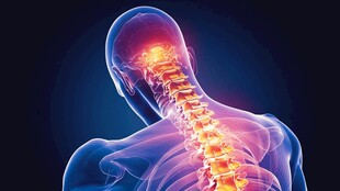
Cervical Spondylosis: Clinical Review for Health Professionals
Introduction
Cervical spondylosis is an umbrella term for age-related degenerative changes of the cervical spine, including intervertebral disc degeneration, osteophyte formation, facet arthropathy, and ligamentous hypertrophy. While radiographic changes are common with aging, a smaller proportion of patients develop symptoms from nerve root or spinal cord compression. Early recognition, appropriate conservative care, and timely surgical referral for progressive myelopathy or refractory radiculopathy are essential to preserve function and prevent irreversible neurologic injury.
Pathophysiology
Degeneration begins with loss of disc hydration and proteoglycan content, causing reduced disc height and abnormal biomechanical loading. Increased stress on facet joints and uncovertebral joints leads to osteophyte formation. Ligamentum flavum hypertrophy and disc protrusion narrow the spinal canal and neural foramina, compressing nerve roots or the cervical spinal cord. Chronic compression can induce ischemia, demyelination, and eventual myelomalacia of the cord.
Clinical Presentation
Symptoms vary by the structures involved:
– Axial neck pain and stiffness: Common, often exacerbated by sustained posture or motion.
– Cervical radiculopathy: Unilateral neck and arm pain with dermatomal paresthesia, sensory loss, or focal motor weakness (commonly C6–C7).
– Cervical myelopathy: Gait disturbance, hand clumsiness, hyperreflexia, Hoffmann sign, Babinski sign, and bladder or bowel dysfunction in advanced cases.
– Vertebrobasilar symptoms: Dizziness, visual disturbance, or syncope are less common and should prompt vascular evaluation.
Red flags include rapidly progressive neurologic deficit, intractable pain, constitutional symptoms, or signs suggesting infection or malignancy.
Diagnostic Evaluation
- History and neurological exam: Document onset, progression, radicular distribution, motor deficits, gait abnormalities, and sphincter symptoms. Use objective scales (Nurick, mJOA) when assessing myelopathy severity.
- Imaging: Plain radiographs for alignment and dynamic instability (flexion/extension views). MRI is the investigation of choice to evaluate cord compression, disc pathology, and signal changes within the cord. CT is valuable for bony detail and preoperative planning.
- Electrodiagnostics: Nerve conduction studies and EMG help differentiate radiculopathy from peripheral neuropathy when the clinical picture is unclear.
Conservative Management
Most patients with mild radiculopathy or axial neck pain respond to non-operative treatment:
– Activity modification and ergonomic advice: Avoid aggravating positions; optimize workstation setup.
– Analgesia and short-course anti-inflammatories: NSAIDs, acetaminophen; consider neuropathic agents (gabapentin, duloxetine) for radicular pain.
– Physical therapy: Supervised programs focusing on cervical stabilization, postural correction, and graded mobilization.
– Cervical traction and collars: Short-term use may relieve symptoms but have limited long-term benefit. Soft collars are used sparingly to reduce pain, not long-term immobilization.
– Epidural steroid injection: Targeted transforaminal or interlaminar injections can be effective for radicular pain when conservative measures fail; benefits are often temporary and used as an adjunct to rehabilitation.
Conservative treatment duration is typically 6–12 weeks unless neurologic deficits progress.
Indications for Surgical Referral
Refer to spine surgery when there is:
– Progressive or significant cervical myelopathy (gait impairment, hand dysfunction).
– Severe, refractory radicular pain with corresponding structural pathology on imaging.
– Radiographic instability, significant deformity, or large central stenosis causing cord compression.
Surgical Options and Considerations
Surgical goals are decompression of neural elements, stabilization when required, and restoration of alignment:
– Anterior Cervical Discectomy and Fusion (ACDF): Effective for focal disc pathology and foraminal stenosis; allows direct decompression and restoration of disc height.
– Cervical disc arthroplasty: Motion-preserving alternative for select single-level disease without facet arthropathy.
– Posterior decompression (laminoplasty/laminectomy ± fusion): Preferred for multilevel central stenosis or ossification of the posterior longitudinal ligament when posterior elements must be preserved or when anterior access is less favorable.
– Instrumentation and fusion: Indicated for instability, deformity, or multilevel reconstructions.
Risk-benefit discussion should include dysphagia, adjacent segment disease, infection, and rare neurologic deterioration.
Perioperative Care (Key Points)
- Preoperative optimization: Address modifiable risks—smoking cessation, glycemic control, nutritional status, and bone health (vitamin D, calcium, DEXA when indicated). Document baseline neurologic status and discuss expectations.
- Pulmonary preparation: Teach incentive spirometry for patients with reduced pulmonary reserve or thoracic involvement.
- Intraoperative monitoring: Consider somatosensory and motor evoked potential monitoring for multilevel decompressions or deformity corrections.
- Postoperative management: Frequent neurologic checks, wound and drain monitoring, early mobilization with cervical support as directed, thromboprophylaxis per institutional protocol, and multimodal analgesia. Swallowing assessment and attention to airway in anterior approaches are critical in early postoperative period.
Rehabilitation and Follow-Up
- Early mobilization: Initiate gentle range-of-motion and progressive strengthening as permitted by the surgical plan.
- Long-term follow-up: Serial clinical and radiographic assessments to monitor fusion status, hardware integrity, and adjacent segment disease. Use standard outcome measures (NDI, mJOA) to track recovery.
Practical Clinical Pearls
- Use objective myelopathy scales to guide timing of surgery—early intervention for deteriorating myelopathy improves outcomes.
- Epidural steroid injections may delay but do not replace surgery for progressive cord compression.
- Document neurologic findings thoroughly before and after interventions to detect changes promptly.
Conclusion
Cervical spondylosis spans a spectrum from asymptomatic radiographic change to progressive myelopathy. Management requires careful clinical assessment, appropriate imaging, evidence-based conservative care, and timely surgical referral for neurologic compromise. A multidisciplinary approach—incorporating neurosurgery/orthopedics, physiotherapy, pain management, and primary care—optimizes patient outcomes and preserves neurologic function.


