X‑Ray Examination Methods: Principles, Clinical Utility, and Modern Evolution
1. Overview
X‑ray imaging encompasses multiple acquisition strategies tailored to anatomical detail, functional assessment, and diagnostic specificity. Traditional methods—fluoroscopy, projection radiography, and contrast studies—have progressively integrated digital detectors, dose optimization, and advanced post-processing. Specialized examinations extend capability for problem-solving in complex anatomical or physiological scenarios.
- 1. Overview
- 2. Core Modalities
- 3. Fluoroscopy
- 4. Projection Radiography (Plain Films)
- 5. Contrast-Enhanced Conventional Studies
- 6. Specialized X‑Ray Techniques
- 7. Radiation Dose & Safety
- 8. Image Quality Metrics
- 9. Workflow & Documentation
- 10. Quality Improvement Opportunities
- 11. Integration with Advanced Modalities
- 12. Future Directions
- 13. Key Takeaways
2. Core Modalities
| Modality | Primary Purpose | Real-Time? | Typical Use Cases | Key Strengths | Key Limitations |
|———-|—————–|———–|——————|—————|—————–|
| Fluoroscopy | Dynamic visualization | Yes | GI motility, swallowing studies, interventional guidance | Temporal resolution, multi-position assessment | Lower spatial resolution, higher cumulative dose potential |
| Projection Radiography | High-resolution static anatomy | No | Chest, skeletal trauma, lung pathology, line/tube placement | Wide availability, low cost, permanent record | Limited soft-tissue contrast, no intrinsic function info |
| Contrast Radiography | Morphology + function of hollow/vascular structures | Often | Esophagram, cystourethrogram, IV urography (historic), sinography | Enhances delineation of lumens, patency, leaks | Contrast reactions, prep requirements, evolving CT/MR replacement |
| Specialized X‑Ray Techniques | Problem-solving / enhanced detail | Variable | Tomography (historic), magnification views, soft tissue, motion analysis | Targeted visualization, micro-detail | Narrow indications, replaced by CT/MR/US in many settings |
3. Fluoroscopy
3.1 Principles
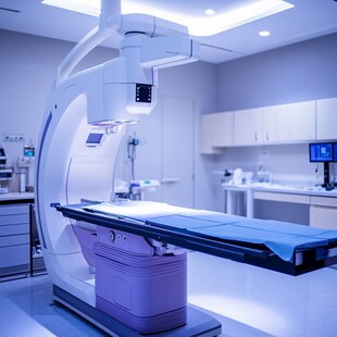
Continuous or pulsed X‑ray beam passes through patient; transmitted photons captured by image intensifier or flat-panel detector producing real-time images.
3.2 Modern Enhancements
- Pulsed fluoroscopy (e.g., 7.5–15 fps) → dose reduction.
- Last-image hold & digital capture → minimize repeat exposures.
- Flat-panel detectors: improved contrast, wider dynamic range vs legacy image intensifiers.
3.3 Clinical Applications
| Category | Examples | Functional Insights |
|———-|———-|——————–|
| Gastrointestinal | Barium swallow, upper GI series, small bowel follow-through | Motility, reflux, obstruction |
| Swallowing / Speech | Videofluoroscopic swallow study | Aspiration, phase timing |
| Musculoskeletal | Stress views, dynamic instability | Ligamentous competence |
| Interventional | Catheter placement, angiographic roadmapping (with DSA adjunct) | Device guidance |
3.4 Advantages & Limitations
| Advantages | Limitations / Risks |
|———–|——————–|
| Multiplanar dynamic assessment | Lower inherent spatial resolution |
| Real-time procedural feedback | Radiation dose accumulation cumulative |
| Functional evaluation (peristalsis, valve motion) | Contrast aspiration risk (swallowing studies) |
| Immediate decision-making | Requires expertise for interpretation |
4. Projection Radiography (Plain Films)
4.1 Physics & Image Formation
- Differential attenuation (photoelectric + Compton) forms contrast.
- Digital detector (computed radiography phosphor plate or direct/indirect flat panel) converts X‑ray signal to electronic data.
4.2 Optimization Parameters
| Factor | Impact | Optimization Strategy |
|——–|——-|———————-|
| kVp | Penetration & contrast | Body part–specific selection; avoid excessive kVp (contrast loss) |
| mAs | Signal (photon flux) & noise | Use AEC; avoid underexposure (quantum mottle) |
| Source–Image Distance (SID) | Magnification & sharpness | Standardized (e.g., 180 cm chest) |
| Collimation | Scatter & dose | Tight collimation improves contrast, reduces dose |
| Grids | Scatter reduction | Use when part >10–12 cm; increase mAs appropriately |
4.3 Clinical Utility
| Region | Key Diagnostic Roles |
|——–|———————|
| Chest | Lung parenchyma overview, cardiomediastinal silhouette, devices |
| Musculoskeletal | Fracture detection, alignment, arthropathy patterns |
| Abdomen | Obstruction signs, calcifications (stones), tube/line localization |
| Mammography (specialized radiography) | Microcalcifications, architectural distortion |
5. Contrast-Enhanced Conventional Studies
5.1 Contrast Agent Classification
| Type | Example | Mechanism | Use |
|——|———|———-|—–|
| Positive (Radio-opaque) | Iodinated, barium sulfate | High-Z attenuation | GI lumen, fistulas, vasculature (DSA) |
| Negative (Radiolucent) | Air, CO2, room air | Low attenuation vs soft tissue | Double-contrast GI, arthrography adjunct |
| Dual (Double-Contrast) | Barium + Gas | Mucosal coating + luminal distention | Detailed gastric/colonic mucosa |
5.2 Delivery Methods
| Method | Description | Examples |
|——–|————|———-|
| Direct Administration | Instilled into target lumen or cavity | Retrograde cystography, fistulography |
| Physiologic Excretion | Systemic administration → organ excretes into lumen | Excretory urography (largely supplanted by CT urography) |
5.3 Indications vs Replacements
| Traditional Study | Current Trend / Replacement | Rationale |
|——————-|—————————|———–|
| Barium enema | CT colonography / colonoscopy | Higher sensitivity; therapeutic capability (colonoscopy) |
| IV urography | CT urography | 3D anatomy & pathology characterization |
| Oral cholecystography | Ultrasound / MRCP | Non-ionizing; superior biliary detail |
| Conventional angiography (diagnostic) | CTA / MRA (for many indications) | Non-invasive, rapid 3D data |
6. Specialized X‑Ray Techniques
| Technique | Principle | Historical/Current Use | Current Status |
|———-|———-|————————|—————|
| Conventional Tomography | Layer imaging via tube & detector motion | Focal lesion localization (renal, lung) | Replaced by CT |
| Magnification Radiography | Small focal spot + increased OID | Microcalcification detail | Limited to mammography & niche uses |
| High-kV Radiography | Elevated kVp for penetration | Large body habitus, spine | Supplanted by protocol optimization |
| Kymography / Motion Recording | Sequential motion capture | Cardiac valve motion (pre-echocardiography) | Obsolete; replaced by echo/MRI |
| Soft X‑Ray | Low kVp & filtration | Surface soft tissue, breast (historical) | Evolved into modern mammography |
| Digital Subtraction Angiography (DSA) | Mask subtraction for vascular contrast | Vascular stenosis, embolization planning | Core interventional tool |
7. Radiation Dose & Safety
| Concept | Plain Film | Fluoroscopy | Mitigation Strategies |
|———|———–|————|———————-|
| Typical Exposure Time | Milliseconds (per exposure) | Seconds–minutes cumulative | Pulsed mode, collimation |
| Dose Monitoring | Dose-area product rarely for plain film | Air kerma & DAP tracked | Real-time dose display |
| Determinants | kVp, mAs, filtration, distance | Fluoro time, frame rate, magnification | Use lowest frame rate, last-image hold |
ALARA principles govern protocol design; justification and optimization are essential.
8. Image Quality Metrics
| Metric | Definition | Relevance |
|——–|———–|———–|
| Spatial Resolution | Ability to distinguish small structures | Microfractures, calcifications |
| Contrast Resolution | Discern differences in tissue densities | Soft tissue detection |
| Noise (Quantum Mottle) | Random pixel intensity variation | Can obscure low-contrast lesions |
| Detective Quantum Efficiency (DQE) | Efficiency of detector converting photons to signal | Dose efficiency trade-offs |
| Modulation Transfer Function (MTF) | Frequency response of system | Predicts fine detail reproduction |
9. Workflow & Documentation
| Step | Radiography | Fluoroscopy |
|——|————|————|
| Order Justification | Clinical indication appropriateness | Same + interventional planning |
| Patient Prep | Remove artifacts (metal) | Fasting (GI), bowel prep (contrast) |
| Acquisition | Standardized projections | Dynamic positioning, contrast administration |
| Recording | DICOM image set archival | Fluoro save frames, dose report |
| Reporting | Structured report supports comparison | Real-time procedural notes + final report |
10. Quality Improvement Opportunities
| Domain | Strategy |
|——–|———|
| Dose Reduction | Protocol audits, AI-driven exposure modulation |
| Repeat Rate Minimization | Technologist education, positioning tools |
| Report Consistency | Structured templates, critical finding flags |
| Turnaround Time | Workflow automation, priority routing |
| Patient Safety | Contrast allergy screening, pregnancy status verification |
11. Integration with Advanced Modalities
Conventional X‑ray remains first-line for skeletal trauma, chest evaluation, line/tube verification, and initial triage. Cross-sectional imaging (CT, MRI, ultrasound) complements or replaces certain legacy studies by providing superior soft-tissue contrast, 3D anatomy, or functional data. Fluoroscopy maintains primacy in interventional radiology, while DSA remains indispensable for endovascular therapy.
12. Future Directions
| Innovation | Potential Impact |
|———–|——————|
| AI Exposure Control | Real-time optimization reduces dose variability |
| Automated Position Recognition | Fewer repeats, standardized projections |
| Spectral (Dual-Layer) Detectors | Material decomposition, contrast dose reduction |
| Edge-Enhancement Algorithms (AI) | Improved diagnostic confidence at lower dose |
| Integrated Dose Registries | Benchmarking & regulatory compliance |
13. Key Takeaways
- Fluoroscopy excels at dynamic functional assessment; radiography at high-resolution structural depiction.
- Contrast studies persist where luminal physiology and direct visualization matter, though many have ceded ground to CT/MR.
- Specialized techniques have largely transitioned to or been replaced by cross-sectional imaging innovations.
- Systematic dose management and quality processes sustain diagnostic yield while minimizing patient risk.
- Emerging AI and spectral detector technologies aim to unify image quality gains with further dose efficiency.
Disclaimer: Educational summary; adhere to institutional radiation safety policies and regional imaging guidelines.


