Scoliosis: Clinical Review for Health Professionals
Introduction
Scoliosis is a three-dimensional deformity of the spine characterized by lateral curvature and axial rotation. It ranges from benign, small curves to progressive deformities that impact cardiopulmonary function, cause chronic pain, and impair quality of life. Timely detection, appropriate conservative management, and judicious surgical referral are central to optimal outcomes.
- Introduction
- Classification and Epidemiology
- Natural History and Clinical Significance
- Clinical Assessment
- Imaging and Measurements
- Non-surgical Management
- Surgical Indications and Options
- Perioperative Considerations and Care (Key points for clinicians)
- Complications and Long-term Follow-up
- Practical Tips for Clinicians
- Conclusion
Classification and Epidemiology
- By age: Infantile (<3 years), juvenile (3–10 years), adolescent (10–18 years), and adult degenerative scoliosis.
- By cause: Idiopathic (≈80% of cases; most common in adolescents), congenital (vertebral malformations), neuromuscular (e.g., cerebral palsy, neuromuscular dystrophy), and degenerative (adult onset).
- Prevalence: Adolescent idiopathic scoliosis (AIS) affects ~2–3% of adolescents; clinically significant curves needing treatment are less common.
Natural History and Clinical Significance
Small curves (<20° Cobb) rarely progress after skeletal maturity. Curves between 20–40° may progress during growth and warrant close surveillance. Curves >45–50° in skeletally immature patients or >50° in adults are more likely to progress and can lead to pain, progressive deformity, and cardiopulmonary compromise when severe.
Clinical Assessment
- History: Onset and progression, pain, neurologic symptoms (radicular pain, weakness), functional limitations, and family history. Screen for red flags: rapid curve progression, nocturnal pain, systemic symptoms.
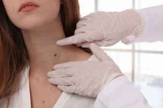
- Physical examination: Adam’s forward bend test for rib prominence or lumbar hump, shoulder/waist asymmetry, limb-length discrepancy, and neurological exam (motor, sensory, reflexes). Document trunk shift and pelvic obliquity.
- Functional assessment: Evaluate respiratory symptoms, exercise tolerance, and impact on activities of daily living.
Imaging and Measurements
- Standing full-spine PA and lateral radiographs: Cobb angle measurement is the standard for quantifying curvature and monitoring progression. Include bending films to assess curve flexibility when planning intervention.
- MRI: Indicated for atypical curves, neurologic deficits, rapid progression, or suspicion of intraspinal pathology (e.g., syrinx, tethered cord).
- EOS or low-dose imaging: Consider for serial monitoring to reduce radiation in adolescents.
Non-surgical Management
- Observation: For skeletally immature patients with small curves (Cobb <20°) or mature patients with stable curves. Monitor with periodic radiographs (every 6–12 months during growth).
- Physical therapy: Targeted exercise programs (Schroth method, core strengthening, postural training) improve trunk muscle balance and may reduce progression in some patients.
- Bracing: Indicated for skeletally immature patients with curves between ~20–45° and documented progression. Effective brace wear (wear time often 16–23 hours/day) can reduce the risk of progression to surgical thresholds. Custom thoracolumbosacral orthoses (TLSO) are most commonly used. Patient adherence and regular follow-up are essential.
- Pain management: Analgesics, activity modification, and multimodal approaches for symptomatic adult degenerative scoliosis.
Surgical Indications and Options
Indications include progressive curves despite bracing, severe deformity (typically Cobb >45–50° in adolescents or symptomatic adult curves), significant pain or neurologic compromise, and trunk imbalance with functional impairment.
– Posterior spinal fusion with segmental instrumentation: The most common approach for AIS; aims to correct deformity and achieve long-term fusion.
– Anterior approaches or anterior-posterior combined procedures: Considered for select curves to improve correction or fusion levels.
– Osteotomies and deformity correction: For rigid or severe deformities; require experienced surgical teams and careful preoperative planning.
– Minimally invasive techniques: Growing use in select adult degenerative cases to reduce morbidity.
Perioperative Considerations and Care (Key points for clinicians)
Preoperative
- Optimization: Correct nutritional deficits, tobacco cessation, and treatment of infection. Evaluate bone health (DEXA) in adults; consider vitamin D and calcium repletion.
- Pulmonary preparation: Respiratory physiotherapy and incentive spirometry for patients with thoracic deformity to reduce postoperative pulmonary complications.
- Neurologic assessment and planning: Document baseline neurologic status; plan for intraoperative neuromonitoring (SSEPs, MEPs) in complex cases.
- Traction and flexibility testing: Suspensory traction or serial casting may be used in rigid curves to improve correction and reduce surgical risk.
Intraoperative
- Blood conservation: Anticipate significant blood loss; use tranexamic acid, cell salvage, and staged procedures when appropriate.
- Neuromonitoring: Continuous SSEPs/MEPs to detect cord compromise early.
- Instrumentation strategy: Choose fusion levels to balance deformity correction with preservation of motion segments and minimize adjacent-segment disease.
Postoperative
- Neurologic checks: Frequent monitoring of motor and sensory function immediately post-op.
- Drain management: Monitor wound drainage volume and character; remove drains per protocol (commonly 24–72 hours).
- Early mobilization: Begin assisted mobilization as permitted (often within 24–48 hours) to reduce pulmonary and thromboembolic complications.
- Wound care and infection surveillance: Monitor for signs of surgical site infection; administer antibiotics per institutional protocol.
- Pain control and rehabilitation: Multimodal analgesia and early initiation of physiotherapy focusing on gait, core strength, and safe activities to prevent recurrence and aid recovery.
Complications and Long-term Follow-up
- Complications: Implant failure, proximal or distal junctional kyphosis, infection, neurologic injury, and pulmonary complications. Discuss risks and realistic outcomes with patients preoperatively.
- Follow-up: Serial radiographic assessment at 6 weeks, 3 months, 6 months, and annually to monitor fusion, implant integrity, and adjacent-segment changes. Long-term rehabilitation and psychosocial support are often necessary, particularly in severe or neuromuscular cases.
Practical Tips for Clinicians
- Screen adolescents with a forward-bend test during routine exams to identify early curves.
- Emphasize adherence to brace wear and structured physiotherapy—these determine non-surgical success.
- Use multidisciplinary teams (orthopedics, neurosurgery, physiotherapy, respiratory therapy, nursing, and social work) for complex deformities and perioperative care.
Conclusion
Scoliosis management balances surveillance, conservative care, and timely surgical intervention when indicated. Clinicians should individualize treatment by patient age, curve magnitude and flexibility, symptom burden, and comorbidities to minimize progression, relieve symptoms, and preserve function.


