Parasitic Infections
Parasitic infection occurs when helminths or protozoa enter the human host and persist, leading to asymptomatic carriage or disease (parasitosis). Understanding lifecycle stages and characteristic clinical patterns is essential for accurate diagnosis and control.
Key Lifecycle Terms
- Infective stage: the developmental form that enters humans to initiate infection (e.g., filariform larvae of Strongyloides, metacercariae of liver flukes, cysts of Giardia).
- Pathogenic stage: the form responsible for tissue damage and clinical manifestations (e.g., Schistosoma eggs inducing granulomas and fibrosis).
- Diagnostic stage: the form typically detected in clinical specimens (e.g., eggs in stool/urine, trophozoites/cysts on O&P, microfilariae in blood).
- Transmissible stage: the form exiting the host or vector that enables spread to new hosts (e.g., malaria gametocytes to mosquitoes; helminth eggs to environment).
Hallmark Clinical–Epidemiologic Patterns
1) Carriers (Asymptomatic Infection)
– Many parasitic infections are silent; infected persons can shed diagnostic/transmissible stages and sustain transmission chains.
2) Chronic Infection
– Without adequate therapy, parasites may persist for months to years. Tissue injury and repair can coexist (e.g., schistosomal egg granulomas alongside periportal fibrosis).
3) Latent/Opportunistic Infection
– Low‑level or latent infection may be undetectable by routine tests and later reactivate with immunosuppression, causing severe disease.
– Examples: Strongyloides stercoralis hyperinfection with corticosteroids; Toxoplasma gondii reactivation in HIV; Cryptosporidium spp. chronic diarrhea in advanced immunodeficiency.
4) Polyparasitism (Multiple Concurrent Infections)
– Concurrent infections with multiple parasites are common and can interact biologically, modifying pathogenicity and clinical expression.
– Interactions may be synergistic or inhibitory; co‑infections can alter susceptibility to other pathogens and response to therapy.
5) Ectopic Parasitism
– Parasites in atypical locations can cause ectopic lesions (diagnostic challenge).
– Examples: intestinal nematodes occasionally entering the urogenital tract or biliary tree; Paragonimus westermani typically pulmonary but may invade CNS; Schistosoma japonicum eggs can embolize to brain or lungs.
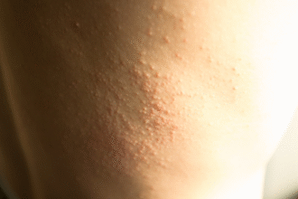
6) Larva Migrans
– Larval stages in non‑definitive hosts migrate through tissues causing inflammation.
– Cutaneous larva migrans: serpiginous pruritic tracks (e.g., Ancylostoma species from contaminated sand/soil).
– Visceral/ocular larva migrans: Toxocara canis/cati larvae causing eosinophilia, hepatic or ocular lesions.
Diagnosis
- History & exposure: travel/residence in endemic areas, freshwater exposure, undercooked meat/fish, barefoot soil contact, animal contact, immunosuppression.
- Labs: CBC for eosinophilia (typical in tissue‑migrating helminths), liver/renal function as indicated.
- Stool/urine Ova & Parasite (O&P): multiple specimens on separate days increase yield; consider concentration techniques.
- Antigen/PCR assays: Giardia, Cryptosporidium, Entamoeba histolytica, malaria NAAT/rapid tests.
- Serology: for tissue parasites not readily detected in stool (e.g., Toxocara, cysticercosis, strongyloidiasis, echinococcosis, schistosomiasis in travelers).
- Imaging: ultrasound/CT/MRI for cystic lesions (echinococcal cysts, neurocysticercosis), biliary obstruction (ascariasis), pulmonary nodules (paragonimiasis).
- Specialized tests: blood films or quantitative buffy coat for malaria; timed microfilariae sampling; duodenal aspirates/agar plate culture for Strongyloides where available.
Management (General Principles)
- Treat confirmed infections with targeted agents and address complications; consider empiric therapy in high‑probability scenarios when diagnostics are limited, following guidelines.
- Common agents: albendazole, mebendazole (many nematodes); ivermectin (strongyloidiasis, onchocerciasis); praziquantel (schistosomiasis, many cestodes/trematodes); nitazoxanide (cryptosporidiosis); metronidazole/tinidazole plus luminal agents for Entamoeba histolytica; artemisinin‑based combinations for malaria (per protocols).
- Corticosteroid use: screen for Strongyloides prior to initiation in at‑risk individuals; give preemptive ivermectin when screening not feasible and risk is high.
- Manage inflammation and mass effect (e.g., steroids in neurocysticercosis per specialist guidance) and surgical/endoscopic interventions as needed (e.g., biliary ascariasis, hydatid cysts—special precautions to avoid spillage/anaphylaxis).
- Coordinate care with infectious disease/tropical medicine specialists for complex or CNS disease.
Prevention
- Water, Sanitation, and Hygiene (WASH): safe drinking water, latrine use, hand hygiene with soap, safe disposal of feces.
- Food safety: cook meat/fish thoroughly; avoid raw/undercooked freshwater fish/crabs; wash produce; safe street‑food practices.
- Environmental/behavioral: footwear to prevent hookworm; avoid skin contact with contaminated freshwater (schistosomiasis); vector control (nets/repellents) for vector‑borne protozoa.
- Animal health: deworm pets; manage stray animals; avoid feeding raw offal to dogs (Echinococcus risk).
- Travel medicine: pre‑travel counseling, chemoprophylaxis for malaria, safe water and food practices.
Red Flags — Seek Urgent Care
- Neurologic symptoms (seizures, focal deficits, severe headache), visual changes.
- Signs of biliary obstruction (jaundice, right upper quadrant pain, fever) or intestinal obstruction.
- Severe anemia, failure to thrive in children, or persistent high fevers.
- Immunocompromised host with new GI, respiratory, or disseminated symptoms.
Educational information only; diagnosis and treatment should follow current parasitology and infectious disease guidelines with specialist input for complicated cases.