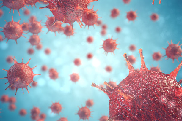
Rabies: How It Spreads in the Body and What It Damages
Rabies is a neurotropic, almost uniformly fatal viral infection once clinical symptoms appear. Understanding its path through the body explains hallmark signs such as hydrophobia, aerophobia, hypersalivation, and autonomic instability. This article summarizes the infection stages, why the virus favors nerves, the immune-evasion tactics it uses, and the key pathological findings.
The Infection Journey: Three Stages
1) Local replication at the entry site (days)
– Entry: Virus enters via broken skin or mucosa, usually through a bite or scratch.
– Local amplification: Initial low-level replication in myocytes and other cells near the wound for several days.
– Nerve entry: Virus accesses nearby peripheral nerves, often at the neuromuscular junction.
2) Rapid centripetal spread to the CNS
– Axonal transport: The virus travels retrograde along peripheral nerve axons toward the dorsal root ganglia, spinal cord, and brain.
– Neurotropism: High replication in the brainstem and cerebellum; limbic and hippocampal structures are commonly involved.
3) Centrifugal dissemination to organs and secretions
– Outflow: From the CNS, the virus spreads along peripheral nerves to many tissues.
– High viral loads: Especially in salivary glands, taste buds, and the olfactory epithelium—facilitating transmission via saliva.
Why Rabies Targets Nerves (Neurotropism) and Evades Immunity
- Neuronal entry and transport: The virus binds receptors at neuromuscular junctions and uses dynein-mediated retrograde axonal transport to reach the CNS.
- Limited neuronal apoptosis: Infected neurons often avoid apoptosis, enabling sustained production and neuron-to-neuron spread.
- Immune privilege in the CNS: Although antigen-specific T cells can enter, antiviral responses are blunted; infiltrating cells may be ineffective or cleared, allowing ongoing replication with relatively modest early inflammation.
Clinical Correlations: How Damage Produces Symptoms
- Brainstem and cranial nerve nuclei (vagus, glossopharyngeal, hypoglossal) involvement drives spasms of respiratory and swallowing muscles, leading to hydrophobia, aerophobia, dysphagia, and dyspnea.
- Autonomic dysfunction from vagal and sympathetic pathway involvement causes hypersalivation, sweating, blood pressure and heart rate instability, and can precipitate arrhythmias or sudden death.
Pathology: What We See Under the Microscope
- Pattern: Acute, diffuse encephalomyelitis with prominent involvement of the hippocampus, brainstem (midbrain, pons, medulla), and cerebellum.
- Gross findings: Congestion, edema, and small hemorrhages may be present.
- Microscopy: Nonspecific neuronal degeneration and inflammatory cell infiltration; perivascular cuffing and microglial nodules can be seen.
- Negri bodies: Eosinophilic cytoplasmic inclusions—aggregates of rabies viral material—classically in hippocampal pyramidal neurons and cerebellar Purkinje cells; typically round/oval, about 3–10 μm, and readily visualized with H&E staining. Presence is supportive but not universal.
Factors That Influence Incubation and Severity
- Inoculum size and virus strain.
- Bite location and innervation density (face and hands have shorter paths to the CNS than distal lower limbs).
- Depth and number of wounds.
- Host factors, including prior vaccination or immune status.
Diagnosis (Brief Overview)
Definitive diagnosis during life relies on laboratory testing (e.g., RT-PCR from saliva, skin biopsy from the nape of the neck, or cerebrospinal fluid; detection of viral antigen). In animals, direct fluorescent antibody testing of brain tissue is standard. Clinical suspicion is driven by compatible exposure plus evolving neurological signs.
Key Takeaways
- Rabies spreads by retrograde axonal transport to the CNS, then outward to peripheral tissues, especially salivary glands.
- The virus’s neurotropism and immune-evasion strategies allow extensive CNS infection with limited early inflammation.
- Classic symptoms map to brainstem and autonomic involvement and predict a grave prognosis once they begin.
- Prevention through timely post-exposure prophylaxis is critical; once symptomatic disease develops, survival is exceedingly rare.
Educational content only. For exposures or symptoms, seek urgent medical care.


